Fluorography of the lungs.
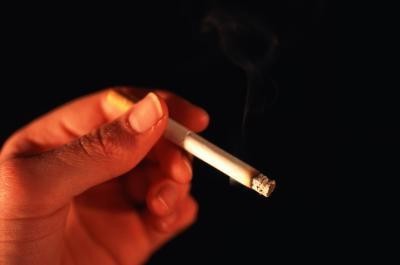
Diseases of the lungs - one of the most frequentproblems, which catches the smoker by surprise. The thing is that the initial changes in the airways occur even after the first cigarette smoked, however, smokers do not notice it.
Fluorography of the lungs is carried out once a yearto all people who have reached the age of 18. If the patient has clinical signs of severe pulmonary disease, but he is not yet 18 years of age, the lung fluorography is performed with the consent of parents, relatives or by collecting a medical commission.
Fluorography is a study with the help ofX-rays that penetrate through the lung tissue and with the help of fluorescent particles transfer the structural pattern of the lungs to the film. Due to the fact that individual tissues and organs absorb x-ray radiation differently, the film is not uniformly colored: a light spot is the heart, and bronchi and bronchioles look lighter. However, the pulmonary tissue should be uniform and homogeneous. With pulmonary diseases on the fluorography will be seen deviation in the form of areas of darkening - increased tissue density or increased airiness - light areas.
Fluorography of the lungs is not always sufficiently informative in comparison with other diagnostic methods, however, this study is ideally suited as a screening diagnostic.
Fluorography of the lungs allows you to suspect any changes in the structure of lung tissue, and then you need to conduct a number of other diagnostic methods for establishing an accurate diagnosis.
Unfortunately, the fluorography of the smoker is not alwayscan fix changes in the lungs, because often they start with the bronchial tree, and not with lung tissue. If you take smokers with an experience of 20 years or more, then, of course, in the picture they can detect specific changes in the lungs, however, they can not 100% talk about nicotine tissue damage.
If we consider smoking as a mechanismprovocation of lung diseases, it is necessary to mention its nonspecificity. When a person absorbs a nicotine suspension, it settles, first of all, on the mucous membranes of the mouth, pharynx, trachea and bronchi, thus causing an inflammatory reaction. That's why smokers after a while begin to cough, and it becomes difficult for them to breathe. The longer a person smokes, the more nicotine is absorbed into the blood, thereby causing toxic damage to organs and systems. But interestingly, with the blood flow, most nicotine particles still settle in the lungs, having a carcinogenic effect.
At this moment on the fluorography you can see the tightness of the lung tissue and reduce its airiness. Cavities filled with pus or other contents may form.
In the picture, one will see an increase in the pulmonary pattern and an expansion of the roots of the lungs, i.e. all signs that the lungs are trying to compensate for this process.
The great advantage of fluorography is that thisthe procedure does not take much time and economic costs are also acceptable. Digital fluorography is now increasingly used. The difference between this method and the standard one is that the image is fixed not on a film, but on a digital medium. It can be viewed, enlarged and brought closer to the desired site. This method is more suitable for a doctor in order to monitor the dynamics of the disease and clarify the diagnosis.
However, despite all the advantages of holdingFluorography in the population, it has one big drawback - radiation load. And let it not be large enough to cause transformation in the body, but still it is not allowed to do fluorography 2-3 times a year.