ECG: the norm of basic indicators
Electrocardiography is an important non-invasivemethod of diagnosis of functional activity of the heart. It is based on the registration of electrical potentials with a contraction of the myocardium, which are displayed on the display or paper.

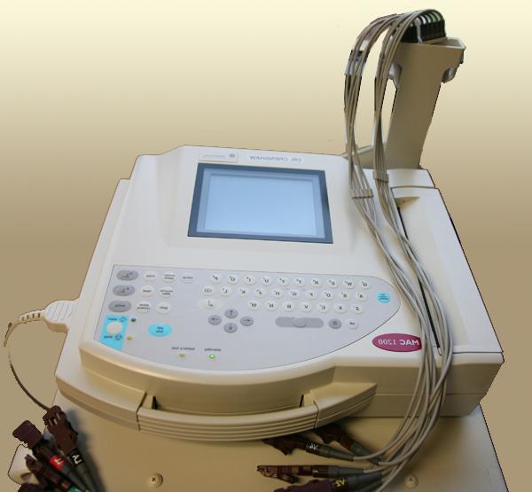
For carrying out an electrocardiogram a special device is used - an electrocardiograph with matching electrodes.
The ECG is normally characterized by the following elements:
1. Teeth, which are denoted by the letters P, Q, R, and also S, T, U.
It should be noted that the tooth P shows how the excitation of the atria passes. Normally, its duration is 0.06-0.1 s, the amplitude is 0.05-2.5 mm.
With the ECG, the norm of the indices is determined taking into accountThe fact that the width of the smallest square on paper where the electrical potentials of the heart are recorded is 0.04 seconds, and the height corresponds to a voltage of 0.1 millivolts.
• Q tooth should be with a duration of <0.03 s and a voltage - <¼ of the amplitude of the tooth R;
• The R tooth should be 0.03-0.04 s and up to 20 mm (in V5 and V6 it can be increased to 26);
• S - normally the duration of this wave reaches 0.03 s, the voltage - <8 in the lead I II, in V1 it can be <25;
• ECG: the tooth norm is T-0.16 s, and the amplitude is <1/2 from the R wave;
• U - the norm is 0,06-16 s, the height of this denticle should be about 2-3 mm.
2. Intervals: PQ (corresponds to the passage of the pulse between the atrium and ventricle), QT, and also RR, ST. The last interval together with the T wave characterizes the repolarization, which passes in the ventricles of the heart;
3. Estimate also the complex QRST, indicating the electric systole of the ventricles.
To record the electrical impulses inmyocardium is used three standard, as well as three reinforced and six thoracic ECG leads. The rate of the results depends on the distance in which the duration and amplitude of the teeth are determined.

PR and ECG amplitude norm is important for correctdiagnosis, because when it increases you can talk about the hypertrophy of the corresponding areas of the heart, which develops in hypertension and certain heart diseases.
It must be said that the ECG is accessible andinformative diagnostic method, but its main disadvantage is the short-term registration of electrical impulses, which spread through the heart muscle. This does not allow us to detect periodic disturbances in the work of the heart. To register such pathologies, monitoring electrocardiography is used, which is performed for 48 hours and gives a more complete picture of the functioning of the myocardium.
In addition, for differentialdiagnosis of organic and functional disorders in the heart can be performed ECG with orthostatic loading or hyperventilation, as well as against the introduction of individual drugs (medicinal tests).